
Products(1) Pixel illuminator-C
Product items
C mount attachable imaging and camera unit
Product name: Pixel
illuminator-C
We supply functionality on microscope
Feature 1: Direct connection to camera port of microscope
•No additional attaching tool is necessary
•No user alignment is necessary
Feature 2: Laser manipulation control on GUI
Application
•Photoconversion•Photoporation (make hole on the cell surface)
•Laser cell screening
•Local heating / IR-LEGO•DNA damage
•Laser marking
Appeal point 1: small and light weight

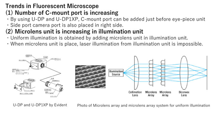
Pixel illuminator is matched to the trends of Fluorescent Microscope
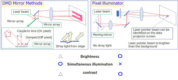
Appeal point 2: High power and high contrast laser illumination
Pixel illuminator is based on laser pointing technology. It is same with that the point of laser pointer is brighter than the presentation screen.
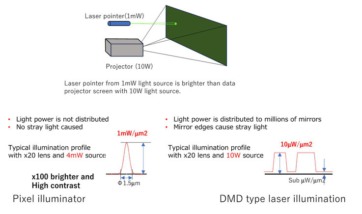
Experimental results of high power and high contrast laser illumination
Pros/Cons against DMD mirror method
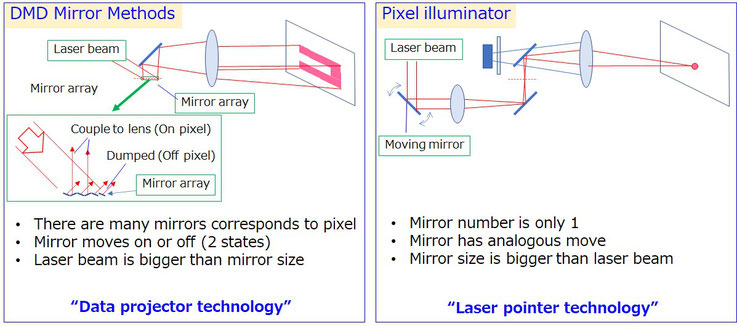
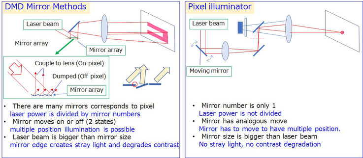
Pros / Cons
Methods of improve of Simultaneous illumination
Appeal point 3: Pixel illuminator can illuminate laser beam with from UV (340nm) to NIR(1550nm).


Methods of improve of Simultaneous illumination (2)
Using two beam scanning system (ordermade)
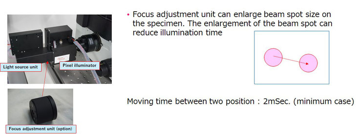
Laser beam illumination profile can be controlled with 1msec resolution.
The beam size enlargement with focus adjustment unit can be applied independently.

Demo Movie 1 CW laser illumination (Selected area illumination )
Boundary selection is set by mouse clicking on image. Laser illumination amount can be adjusted by moving speed and laser intensity
Demo Movie 2 Laser cell selection (Pulsedlaser)
Selected cells are killed by laser illumination in closed chamber. Observation is fluorescent
Demo Movie 3 Object tracking
Laser illumination with image analysis (intensity analysis) is also possible